L3brainsurfaceanatomicalfeatures2png - Correctly label the following anatomical features of the surface of the brain. Label the additional cerebral structures on the right side of the figure Label the regions of gray and white matter in the brain Label the components of the cerebral.
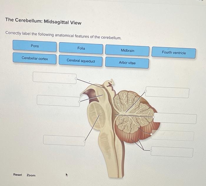
Solved The Cerebellum Midsagittal View Correctly Label The Chegg Com
Located on the lower dorsal aspect of the brain the cerebellum accounts for 11 of the total brain mass.

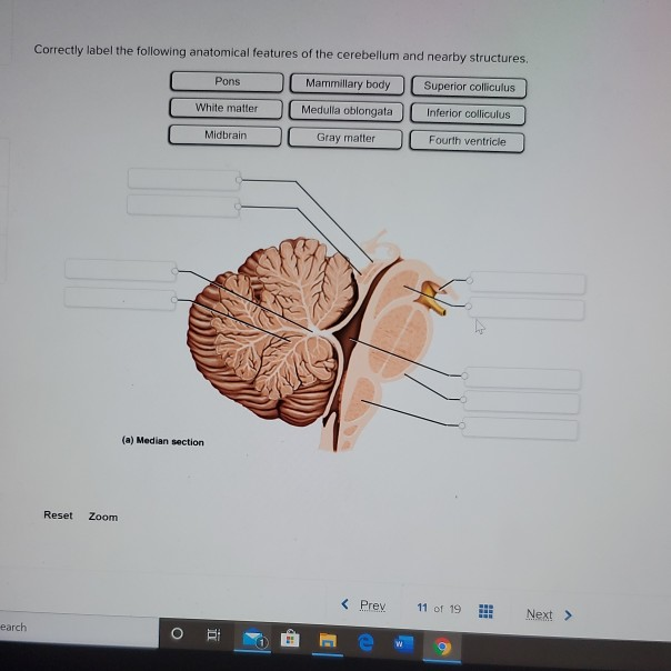
. Correctly label the following anatomical features of the cerebellum and nearby structures Pons Mammillary body Superior colliculus White matter Medulla oblongata Inferior colliculus Midbrain Gray matter Fourth ventricle a Median section Reset Zoom Prev 11 of 19 Next earch O CH. Correctly label the following anatomical features of the surface of the brain Anterior Posterior Spinal cord Brainstem Cerebellum Central sulcus Cerebrum Gyri b Lateral view Temporal lobe Lateral sulcus. Anatomy and Physiology questions and answers.
Identify the indicated region of the brain. Anatomy and Physiology Correctly label the following anatomical features of the cerebellum. Its submitted by giving out in the best field.
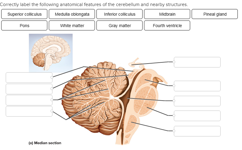
This brain part controls involuntary actions such as breathing heartbeats and digestion. Gray matter and a deeper layer of white matter. Correctly label the following anatomical features of the cerebellum and nearby structures.
It also contains a narrow midline zone called the vermis. Textbook Solutions Expert Tutors Earn. Vertebral arch Spinous Process Nucleus Pulposus Transvere Process Body Vertebral Foramen Anulous Fibrous.
The metencephalon is the upper part of the rhombencephalon or hindbrain. Identify the cranial nerve which controls all but one of the muscles of the palate pharynx and the intrinsic muscles of the larynx. This part of the cerebrum interprets and sorts information from the senses.
Like the cerebral cortex the cerebellum is divided into two hemispheres. Here are a number of highest rated Cerebellar Circuits pictures upon internet. The metencephalon is the upper part of the rhombencephalon or hindbrain.
Each hemisphere exhibits slender transverse parallel folds called folia separated by shallow sulci. We can divide the brain into three major portionsthe cerebrum cerebellum and brainstem. Cerebellum 4 points Lateral sulcus eBook Print References Central sulcus Gyri Brainstem Cerebrum Temporal lobe Spinal cord.
Anatomy and Physiology questions and answers. This part of the nervous system moves messages between the brain and the body. The cerebellum is located dorsal to the pons and medulla and it protrudes under.
We receive this kind of Cerebellar Circuits graphic could possibly be the most trending topic past we allocation it in google help or facebook. The cerebellum consists of right and left cerebellar hemispheres connected by a narrow wormlike bridge called the vermis. Read each description below and determine whether it pertains to the medulla oblongata the pons or the midbrain.
We identified it from honorable source. Each cerebral hemisphere is marked by thick folds called gyri separated by shallow grooves called sulci. Correctly identify the bones and anatomical features of the bones of the skull.
4 Correctly label the following anatomical features of the surface of the brain. Correctly label the following anatomical features of a vertebra. Previous question Next question.
This part of the cerebrum helps us with. Anatomists classify the cerebellum as part of the metencephalon which also includes the pons and all its connections with other parts of the brain travel through the pons. This part of the cerebrum processes messages from the eyes.
Cerebellum Central sulcus Central. Start studying the Chapter 13 Question Set flashcards containing study terms like Identify the cerebral lobes on the left side of the figure. Correctly label the following anatomical features of the cerebellum and nearby structures.
View the full answer. Correctly label the following anatomical features of the surface of the brain. Correctly label the following anatomical features of the cerebellum and nearby structures.
The cerebellum is located at the back of the brain immediately inferior to the occipital and temporal lobes and within the posterior cranial fossaIt is separated from these lobes by the tentorium cerebelli a tough layer of dura mater. Correctly label the following functional regions of the cerebral cortex. Like the cerebral cortex the cerebellum is divided into two hemispheres.
Identify the lobe s of the brain. A set of large folds is by convention used to divide the overall structure into 10 smaller lobules. Frontal Bone Maxilla Mandible Zygomatic Bone.
Prefrontal cortex Wernicke area Motor association area Primary somesthetic cortex Primary motor cortex Somesthetic association area Broca area Visual association area Primary visual cortex. Anterior lobe Anterior Vermis Folia Posterior lobe Posterior Cerebellar hemisphere b. Memorize flashcards and build a practice test to quiz yourself before your exam.
A very deep median groove the longitudinal fissure separates the right. It lies at the same level of and posterior to the pons from which it is separated by the fourth ventricle. Drag each label into the proper location in order to identify the area that would most likely have been affected.
Like the cerebrum the cerebellum has two major hemispheres with an outer cortex made up of gray matter with an inner region of white matter. The cerebellum consists of right and left cerebellar hemispheres connected by a narrow wormlike bridge called the vermis. Correctly label the following anatomical features of the cerebellum.
Identify the indicated regions of the CNS as seen in this anterior view of the brain. Identify the nerve highlighted in the image. Eachhemisphere exhibits slender transverse parallel folds called folia separated by shallow sulci.
Correctly label the following functional regions of the cerebral cortex. The cerebellum has a surface cortex of. 4 Correctly label the following anatomical features of the surface of the brain.
Then click and drag each box into the appropriate category below. Consider a situation where a stroke or mechanical trauma has occurred resulting in damage to one of the areas of the brain indicated in the image.
Solved Correctly Label The Following Anatomical Features Of Chegg Com
Cbio Figures Flashcards Quizlet

Cbio Figures Flashcards Quizlet
Solved Correctly Label The Following Anatomical Features Of Chegg Com


0 Comments